


Recognizing MacTel Masquerades
Real-World Findings Reinforce Encelto’s Role in MacTel Treatment
Macular telangiectasia type 2 (MacTel) is a rare neurodegenerative condition, with an estimated prevalence of 0.045% to 0.1%.1,2 It primarily involves the breakdown of central photoreceptors, retinal pigment epithelium (RPE), and sustentacular Müller cells. The disease itself is associated with genetically driven serine deficiency3 and metabolic accumulation of deoxysphingolipids,4 resulting in slowly progressive loss of juxtafoveal and, subsequently, foveal photoreceptors; juxtafoveal telangiectasia; juxtafoveal intraretinal cavitation; intraretinal crystalline deposits; and, in some cases, progression to subretinal neovascularization (Figures 1 and 2).
Photoreceptor loss in MacTel, measured as ellipsoid zone (EZ) loss on optical coherence tomography, progresses at an average rate of approximately 0.08 mm2 to 0.16 mm2 per year. 5 This slow, progressive neurodegeneration affecting the central macula leads to gradual visual decline, with approximately 15% of eyes losing 15 letters or more (equivalent to 3 Snellen lines) over 5 years and an average best-corrected visual acuity loss of 1 to 2.3 letters per year.6
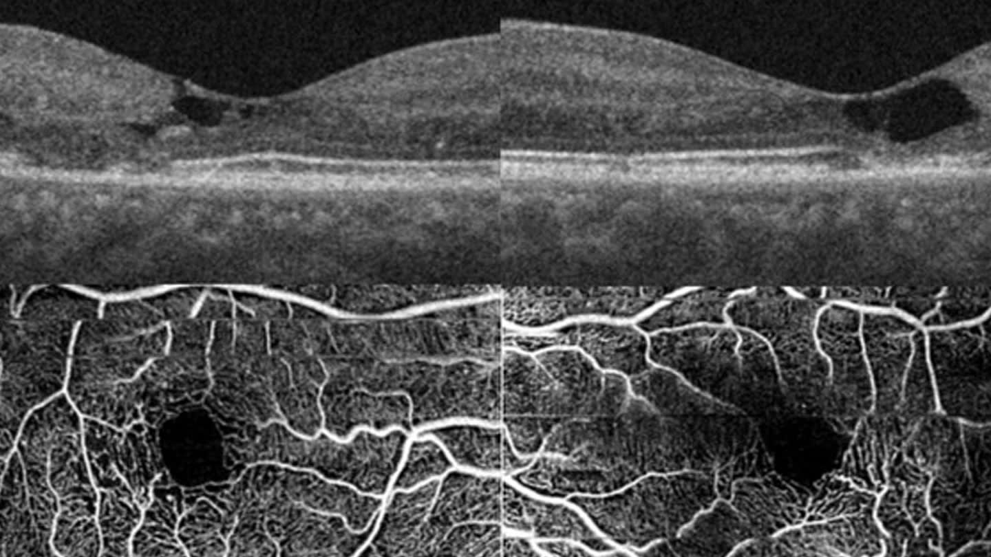
Figure 1. (Top) Optical coherence tomography (OCT) of the right and left macula, respectively, showing temporal juxtafoveal inner retinal cavitation with ellipsoid zone disruption. (Bottom) OCT angiography of the right and left macula, respectively, showing bilateral temporal juxtafoveal macular telangiectasia.
Ciliary neurotrophic factor (CNTF) is a neuroprotective cytokine that may act through enhancement of cellular glycolytic and anabolic pathways and restoration of the antioxidant glutathione. Its neuroprotective effects were first demonstrated in animal models of retinal degeneration in 1992.7,8 Given this neuroprotective role, CNTF has been studied in retinal degenerative diseases.
Continuous delivery of intravitreal CNTF is possible via encapsulated cell technology: an intraocular implant containing approximately 200,000 to 440,000 engineered live human RPE cells enclosed within a semipermeable fibrous membrane. The surgically implanted, intravitreal, scleral-anchored revakinagene taroretcel-lwey implant (Encelto; Neurotech Pharmaceuticals), formerly known as NT-501, is positioned at the pars plana and allows bidirectional exchange—nutrient inflow to sustain the RPE cells and CNTF outflow to the retina—while protecting the RPE cells from host immune rejection.9 Retrospective analysis of explanted devices has demonstrated sustained production of bioactive CNTF for up to 14.5 years, suggesting the potential for long-term photoreceptor and Müller cell preservation.10
In phase 1 and 2 clinical trials in MacTel, revakinagene demonstrated efficacy in reducing photoreceptor loss.11-14 In the phase 3 randomized, masked, sham-controlled trials (NTMT-03A and NTMT-03B) that led to revakinagene becoming the first FDA-approved treatment for MacTel in March 2025, the mean rate of photoreceptor loss, the primary outcome measured by EZ area, was reduced at 24 months by 55% (P<.001) and 31% (P<.02), respectively.15
Although progressive EZ area loss has been shown to correlate with reduced visual function in MacTel,6 secondary functional outcomes in these trials were inconsistent with regard to reading speed, visual sensitivity, and visual acuity. However, pooled data from 4 clinical trials have shown that NT-501 does have a positive impact on these functional outcomes.16 Serious adverse events occurred in 5% of patients and were primarily related to the surgical procedure rather than the therapeutic agent itself; implant extrusion occurred in 0.9%. Dark adaptation, which is classically affected in MacTel, was delayed in 17% to 24% of treated patients compared with 0% to 2% of sham controls over the 2-year study. Miosis was observed in 14% to 17% of treated patients and in none of the controls.15
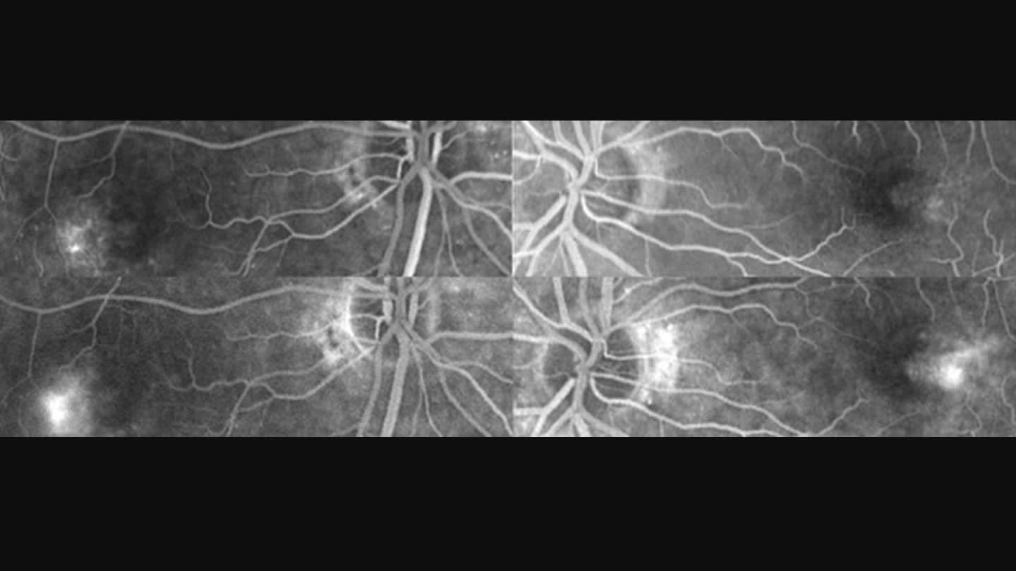
Figure 2. (Top) Early-phase fluorescein angiography of the right and left macula, respectively, in the same patient shown in Figure 1, demonstrating hyperfluorescence of bilateral temporal juxtafoveal macular telangiectasia. (Bottom) Late-phase images show leakage from the telangiectasias and staining corresponding to areas of ellipsoid zone disruption on optical coherence tomography.
Revakinagene has been studied in other neurodegenerative ocular diseases, including inherited retinal dystrophies (retinitis pigmentosa, Usher syndrome, choroideremia, and CNGB3-associated achromatopsia), optic neuropathies such as glaucoma and ischemic optic neuropathy, and geographic atrophy (GA) in age-related macular degeneration. Interest remains in its potential role in GA, for which current therapies are limited to high-burden, indefinite intravitreal complement inhibition. Gene therapies remain investigational, and photobiomodulation, although promising in early studies such as LIGHTSITE, requires further validation.
In 2011, a phase 2 proof-of-concept, multicenter, prospective, double-masked, sham-controlled study of revakinagene in GA resulted in a dose-dependent increase in retinal thickness at 1 year but did not demonstrate efficacy in slowing GA progression.17 However, a single 12-year longitudinal case report suggested slower progression of macular and peripapillary atrophy in the treated eye compared with the untreated eye.18 Despite these efforts, MacTel remains the only clinical condition for which efficacy evidence has supported FDA approval of revakinagene to date.
The concern, of course, is cost. The list price for revakinagene is approximately $250,000—substantial, although lower than that of other landmark gene-based and cell-based therapies such as Lenmeldy ($4.25 million) for metachromatic leukodystrophy, Hemgenix ($3.5 million) for hemophilia B, Zynteglo ($2.8 million) for β-thalassemia, and voretigene neparvovec (Luxturna; $850,000) for biallelic RPE65 mutation-associated retinal dystrophy.
In retinal disease, quality-adjusted life-year (QALY) gains are typically driven by preservation of visual acuity, with associated maintenance of reading ability and sustained independence in activities of daily living. In the MacTel revakinagene trials, the discordance between structural preservation and measurable visual function introduces uncertainty in estimating QALY gains in this slowly progressive disease, particularly when considered against the commonly accepted willingness-to-pay threshold of $150,000 per QALY. However, this assessment may differ for certain subgroups, such as younger patients, those with more rapid structural progression, or those with high visual demands.
Given the typically slow progression of vision loss in MacTel, a critical question emerges: Does the balance of cost and procedural risk justify the structural benefit observed with revakinagene in a given patient with MacTel? Also, what promise does revakinagene hold for other neurodegenerative diseases, such as GA, in the future?
We are fortunate to have input from Martin Friedlander, MD, PhD, professor in the department of cellular and molecular biology, Scripps Research; attending physician, division of ophthalmology, Scripps Clinic; and president, the Lowy Medical Research Institute, La Jolla, California; and Kristin Raming, MD, resident, department of ophthalmology at University Hospital of Bonn, Germany.
Making the Case for Revakinagene
Martin Friedlander, MD, PhD
Kristin Raming, MD
Macular telangiectasia type 2 (MacTel) is a neurovasculoglial degenerative disease characterized by progressive loss of macular function.19 As the disease progresses and involves the foveal center, it results in a significant decline in visual acuity and reading ability and may ultimately progress to legal blindness (20/200 or worse).20 Until recently, no causal or disease-modifying treatment options were available. The emergence of revakinagene taroretcel-lwey represents the first effective therapy targeting the underlying neurodegeneration using a novel encapsulated cell therapy to deliver CNTF.21 A recent phase 3 trial demonstrated the safety and efficacy of revakinagene, with significant slowing of retinal neurodegeneration, including reduced photoreceptor loss and stabilization of macular structure.15
Early Intervention Prevents Irreversible Damage
In earlier disease stages, relative foveal preservation of the EZ on optical coherence tomography may still be present, meaning central vision has not yet been irreversibly lost. During this window, treatment with revakinagene can delay progression toward foveal involvement, thereby preserving central vision for a longer period. With increased awareness of the disease and the availability of enhanced multimodal imaging, patients with MacTel are being diagnosed at earlier stages.22,23 Emerging evidence suggests that revakinagene may be most effective when administered early in the disease course. However, patients with very early disease fell below the inclusion criterion for the phase 3 clinical trial (>0.16 mm2 EZ loss), and the therapeutic effect in this subgroup therefore remains to be fully evaluated. Nevertheless, it is reasonable to assume that eyes with early findings may have a greater treatment response and improved preservation of visual function.
Revakinagene Provides Functional as Well as Structural Benefits
Recent pooled analyses of phase 1, 2, and 3 clinical trial data have shown functional benefits of revakinagene treatment in patients with MacTel.24,25 In the pooled analysis, revakinagene conferred both anatomic and visual function benefits across 3 randomized, sham-controlled studies. Relative to sham, NT-501 demonstrated preservation of anatomy as well as a 36% reduction in photoreceptor loss. With regard to preservation of function, there was a 68% reduction in reading speed loss, a 35% reduction in retinal sensitivity loss, and a 36.2% reduction in the rate of EZ area loss over 24 months.
Long-Term Cost Savings Despite High Initial Costs
Although revakinagene is designed as a one-time therapy (approximately $250,000), comparable retinal treatments involve substantial ongoing costs:
-
Pegcetacoplan (Syfovre; Apellis/Biogen): approximately $25,000 per year with monthly injections
-
Anti-VEGF therapies (eg, ranibizumab, aflibercept): approximately $15,000 to $30,000 or more per year
-
Voretigene neparvovec (Luxturna): approximately $850,000 as a one-time therapy
In this context, revakinagene offers a favorable long-term cost profile compared with chronic treatment strategies.
Rare (Orphan) Disease
The total cost of developing a drug and taking it through the regulatory process averages $350 million to $2.6 billion. Factoring in the time cost of money—the returns investors forgo during the 10-year to 15-year development timeline—and the costs of failed drugs raises the total average estimate to $1.3 billion to $2.6 billion. Recovering these costs is considerably easier for diseases with a high prevalence than for rare diseases. MacTel is a rare disease (prevalence, 0.045% to 0.1%), which justifies special regulatory and reimbursement considerations. High-cost therapies are commonly accepted in such settings because of limited patient populations and a lack of alternatives.
Significant Patient Benefit
Delaying vision loss has substantial clinical and real-world relevance, including:
-
Preservation of reading ability, daily functioning, and the ability to drive;
-
Maintenance of independence and quality of life; and
-
Delay of central visual loss, as measured, for example, by microperimetry.
These effects are not only statistically significant but also directly meaningful for patients.
Socioeconomic Value
It is important to note that patients with MacTel are predominantly of working age, meaning that disease-related visual impairment and treatment burden have a particularly significant impact on professional activity and productivity.26 A delay in visual decline may also lead to:
-
Reduced need for caregiving and assistance;
-
Lower risk of secondary complications, including falls and depression; and
-
Decreased long-term health care and societal costs.
Thus, despite high upfront costs, treatment with revakinagene provides significant value for both the individual and society and consists of a single surgical implantation with only limited follow-up visits. In contrast, intravitreal injection therapies require repeated injections, each involving monitoring, mydriasis, risk of infection, and temporary absence from work. Frequent clinic visits represent a substantial treatment burden for visually impaired patients. This results in a substantially greater treatment burden compared with a one-time approach.
Conclusion
High-cost therapies have consistently been approved for conditions with significant unmet medical need, particularly when irreversible loss of function, such as blindness, is imminent. Decisions are based not only on clinical efficacy but also on prevention of long-term societal costs and preservation of patient autonomy. Therefore, it is our opinion that the benefit of revakinagene is worth the premium cost. RP



References
1. Klein R, Blodi BA, Meuer SM, Myers CE, Chew EY, Klein BE. The prevalence of macular telangiectasia type 2 in the Beaver Dam eye study. Am J Ophthalmol. 2010;150(1):55-62.e2. doi:10.1016/j.ajo.2010.02.013
2. Aung KZ, Wickremasinghe SS, Makeyeva G, Robman L, Guymer RH. The prevalence estimates of macular telangiectasia type 2: the Melbourne Collaborative Cohort Study. Retina. 2010;30(3):473-478. doi:10.1097/IAE.0b013e3181bd2c71
3. Bonelli R, Ansell BRE, Lotta L, et al. Genetic disruption of serine biosynthesis is a key driver of macular telangiectasia type 2 aetiology and progression. Genome Med. 2021;13(1):39. doi:10.1186/s13073-021-00848-4
4. Gantner ML, Eade K, Wallace M, et al. Serine and lipid metabolism in macular disease and peripheral neuropathy. N Engl J Med. 2019;381(15):1422-1433. doi:10.1056/NEJMoa1815111
5. Heeren TFC, Kitka D, Florea D, et al. Longitudinal correlation of ellipsoid zone loss and functional loss in macular telangiectasia type 2. Retina. 2018;38 Suppl 1(Suppl 1):S20-S26. doi:10.1097/IAE.0000000000001715
6. Peto T, Heeren TFC, Clemons TE, et al. Correlation of clinical and structural progression with visual acuity loss in macular telangiectasia type 2: MacTel project report no. 6. Retina. 2018;38 Suppl 1(Suppl 1):S8-S13. doi:10.1097/IAE.0000000000001697
7. LaVail MM, Unoki K, Yasumura D, Matthes MT, Yancopoulos GD, Steinberg RH. Multiple growth factors, cytokines, and neurotrophins rescue photoreceptors from the damaging effects of constant light. Proc Natl Acad Sci U S A. 1992;89(23):11249-11253. doi:10.1073/pnas.89.23.11249
8. Wen R, Tao W, Li Y, Sieving PA. CNTF and retina. Prog Retin Eye Res. 2012;31(2):136-151. doi:10.1016/j.preteyeres.2011.11.005
9. Sieving PA, Caruso RC, Tao W, et al. Ciliary neurotrophic factor (CNTF) for human retinal degeneration: phase I trial of CNTF delivered by encapsulated cell intraocular implants. Proc Natl Acad Sci U S A. 2006;103(10):3896-3901. doi:10.1073/pnas.0600236103
10. Kauper K, Nystuen A, Orecchio L, et al. Long-term durability of ciliary neurotrophic factor–releasing revakinagene taroretcel-lwey in individuals with retinal degenerative disorders. Invest Ophthalmol Vis Sci. 2025;66(11):3. doi:10.1167/iovs.66.11.3
11. Chew EY, Clemons TE, Peto T, et al. Ciliary neurotrophic factor for macular telangiectasia type 2: results from a phase 1 safety trial. Am J Ophthalmol. 2015;159(4):659-666.e1. doi:10.1016/j.ajo.2014.12.013
12. Chew EY, Clemons TE, Jaffe GJ, et al. Effect of ciliary neurotrophic factor on retinal neurodegeneration in patients with macular telangiectasia type 2: a randomized clinical trial. Ophthalmology. 2019;126(4):540-549. doi:10.1016/j.ophtha.2018.09.041
13. Yan J. Phase 3, multicenter, randomized, sham-controlled studies of the efficacy and safety of revakinagene taroretcel (NT-501) in macular telangiectasia type 2. Presented at: The Retina Society annual meeting; Lisbon, Portugal; September 2024.
14. Singerman LJ, Hubschman JP, Friedlander M, et al. Results from a phase I extension study of ciliary neurotrophic factor in patients with macular telangiectasia type 2. Ophthalmol Sci. 2025;6(2):101009. doi:10.1016/j.xops.2025.101009
15. Chew EY, Gillies M, Jaffe GJ, et al. Cell-based ciliary neurotrophic factor therapy for macular telangiectasia type 2. NEJM Evid. 2025;4(8):EVIDoa2400481. doi:10.1056/EVIDoa2400481
16. Goldberg R, Manning D, Yankey J, et al. Pooled analyses of anatomic and visual function data of revakinagene taroretcel-lwey in participants with macular telangiectasia type 2. Ophthalmology Retina; paper in press, 2026.
17. Zhang K, Hopkins JJ, Heier JS, et al. Ciliary neurotrophic factor delivered by encapsulated cell intraocular implants for treatment of geographic atrophy in age-related macular degeneration. Proc Natl Acad Sci U S A. 2011;108(15):6241-6245. doi:10.1073/pnas.1018987108
18. Shiromani S, Leef D, Jain N, Yan J. Ciliary neurotrophic factor implant in geographic atrophy from age-related macular degeneration: a long-term case study. Retin Cases Brief Rep. 2026;20(2):208-213. doi:10.1097/ICB.0000000000001725
19. Charbel Issa P, Gillies MC, Chew EY, et al. Macular telangiectasia type 2. Prog Retin Eye Res. 2013;34:49-77. doi:10.1016/j.preteyeres.2012.11.002
20. Heeren TFC, Chew EY, Clemons T, et al. Macular telangiectasia type 2: visual acuity, disease end stage, and the MacTel area: MacTel Project report number 8. Ophthalmology. 2020;127(11):1539-1548. doi:10.1016/j.ophtha.2020.03.040
21. Nystuen A, Gonzalez-Lopez E, Kauper KA, Eade K, Aaberg TM Jr. Neuroprotective properties of ciliary neurotrophic factor in the retina for the treatment of macular telangiectasia type 2. Cytokine Growth Factor Rev. 2025;84:12-19. doi:10.1016/j.cytogfr.2025.06.005
22. Heeren TF, Holz FG, Charbel Issa P. First symptoms and their age of onset in macular telangiectasia type 2. Retina. 2014;34(5):916-919. doi:10.1097/IAE.0000000000000082
23. Raming K, Rodriguez Garcia JL, Steffens N, et al. Frequency of misdiagnoses and associated risk factors in macular telangiectasia type 2. Retina. 2026;46(2):301-308. doi:10.1097/IAE.0000000000004681
24. Clark WL, Bitar M, Manning DC, et al; the MacTel study investigators. Pooled functionality data of revakinagene taroretcel in patients with macular telangiectasia type 2. Presented at: Hawaiian Eye and Retina 2025; Kauai, Hawaii; January 18-24, 2025.
25. Aaberg T, Goldberg RA, Manning DC, et al; the MacTel Study Group. Pooled functionality data of revakinagene taroretcel-lwey in participants with macular telangiectasia type 2. Submitted for publication.
26. Clemons TE, Gillies MC, Chew EY, et al. The National Eye Institute Visual Function Questionnaire in the macular telangiectasia (MacTel) project. Invest Ophthalmol Vis Sci. 2008;49(10):4340-4346. doi:10.1167/iovs.08-1749