






New Drug Therapies for Benign Retinal Tumors
Despite excellent local control of uveal melanoma, nearly 40% of patients develop metastatic disease over the long term.1 Molecular testing of tumor DNA and RNA has become integrated into clinical practice by stratifying patients into low-risk and high-risk categories for metastasis based on characteristic chromosomal and transcriptional signatures.2 For example, gene expression profiling (GEP) is broken down into class 1 and 2 with ascending risk of metastasis.3
However, DNA and RNA are indirect measures of protein, the effector of cellular physiology, because posttranscriptional and posttranslational modifications can alter protein abundance and downstream signaling independent of transcript levels.4 Although effective, diagnostic and prognostic biopsy requires direct tumor sampling, which is relatively invasive, carrying risk of vitreous hemorrhage, retinal detachment, and sampling bias from tumor heterogeneity.5 This suggests a need for less-invasive methods.
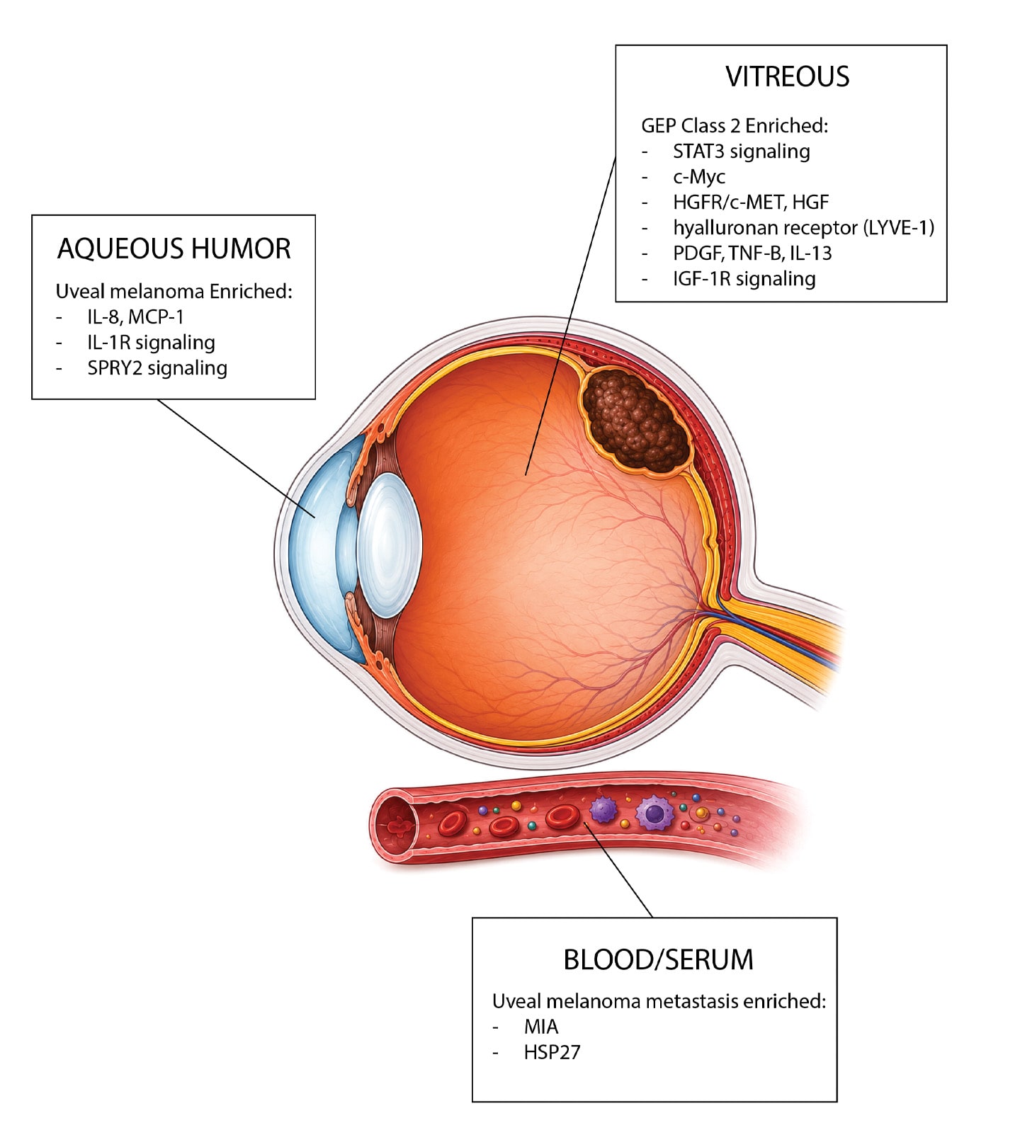
Figure 1. Biomarkers identified in aqueous humor, vitreous, and blood/serum highlight potential applications of proteomics for tumor characterization, prognostication, and metastasis detection.
Proteomics offers a complementary approach by identifying the proteins that govern cellular behavior. Large-scale and unbiased interrogation of the proteome, including posttranslational modifications, can be performed using mass spectrometry coupled with liquid chromatography.4 Smaller-scale targeted approaches can also be used, such as multiplexed enzyme-linked immunosorbent assays (ELISAs).4 As with gene expression changes, characterizing the proteomic alterations that accompany uveal melanoma could assist with diagnosis and prognostication and help identify novel therapeutic targets.
Blood and Serum
Blood-based proteomics has long been used in oncology to detect metastasis and monitor treatment response because of its minimally invasive nature, exemplified by prostate-specific antigen (PSA) testing in prostate cancer.6 In uveal melanoma, early proteomic work focused on cell lines, identifying overexpressed cell cycle genes such as cyclin D1 and cyclin E compared to normal melanocytes.7 Proteomic comparison of a primary uveal melanoma cell line with 2 metastasis-derived cell lines from the same patient identified increased HSP27 expression in metastatic cells.8 Interestingly, subsequent immunohistochemical analysis of primary tumors found reduced HSP27 expression to correlate with monosomy 3 and worse survival, suggesting HSP27 may play distinct roles in the primary tumor microenvironment and established metastases.9 The secretome has similarly been compared between uveal melanoma cells and normal melanocytes to identify candidate systemic biomarkers, including melanoma inhibitory activity (MIA), a protein implicated in the detachment of melanoma cells from the extracellular matrix.10
Moving beyond cell culture, targeted serum proteomics for MIA and related markers in patients with and without metastatic disease demonstrated better sensitivity than conventional liver function tests.11,12 A separate group examined a 49-protein serum panel and achieved 83% sensitivity and 100% specificity for distinguishing uveal melanoma patients from healthy controls. Importantly, these peptides normalized following treatment, suggesting potential utility for both diagnosis and longitudinal monitoring of tumor burden during treatment.13
Aqueous Humor Proteomics
A central challenge of systemic proteomics is that circulating protein profiles represent a dynamic composite of proteins derived from numerous organ systems, creating notable background noise that can obscure disease-specific biomarkers. Ophthalmic diseases, however, are uniquely positioned to benefit from a proteomic approach given the accessibility of the eye for sampling from discrete compartments, such as the anterior chamber and the vitreous cavity. These compartments can be sampled through minimally invasive procedures for diagnosis and longitudinal monitoring, improving the signal-to-noise ratio compared with systemic sampling.14 The anterior chamber is highly accessible, and nearly 90% of vitreous proteins are detectable in aqueous humor, indicating that anterior-chamber sampling can offer insight into posterior segment pathology.15
Even so, ophthalmic proteomic studies are challenged by biologic variability, complex tissue microenvironments, limited ocular sample volumes, and relatively small patient cohorts. Nevertheless, DNA-aptamer–based assays can now quantify nearly 6,000 proteins from as little as 50 μL of aqueous humor.15 An additional workaround is performing unbiased proteomics on small discovery cohorts followed by targeted validation, such as cytokine arrays, in larger populations. Another promising advance is TEMPO (Tracing Expression of Multiple Protein Origins), which integrates proteomics, artificial intelligence (AI), and single-cell transcriptomics to assign aqueous humor proteins to their cellular origins, generating cell-type-specific signatures from a minimally invasive aqueous humor sample.14,15
Aqueous humor proteomics may have diagnostic potential. Targeted profiling of immune mediators in aqueous humor from eyes with uveal melanoma vs benign pigmented lesions showed enrichment of proinflammatory and proangiogenic mediators, such as IL-8 and MCP-1.16
Beyond diagnosis, aqueous humor proteomics may also have prognostic applications. One study with 90 enucleated uveal melanoma eyes evaluated 92 preselected inflammation-related proteins and identified 3 clusters that correlated with tumor size, monosomy 3, gain of chromosome 8q, and loss of BAP1 protein expression. Interestingly, the worst-prognosis cluster was enriched for apoptosis-related proteins.17
A separate study of 20 uveal melanoma aqueous humor samples compared proteomic signatures based on GEP status and found IL-1 receptor and SPRY2 signaling to be potential upstream regulators of the differentially expressed proteins (DEPs).18 All GEP class 2 cases were correctly identified by DEP clustering, whereas 3 GEP class 1 cases were discordant. None of the DEPs overlapped with the 15-gene panel used for commercial GEP testing, reinforcing that the proteome can offer insights distinct from the tumor transcriptome. Additionally, 3 tumors under 2.5 mm, which is normally considered too small to biopsy safely, yielded meaningful aqueous humor proteomic signatures.18
Collectively, these studies demonstrate that aqueous humor proteomics complements conventional GEP by providing independent biologic information through less invasive sampling, which offers the opportunity to perform repeated sampling for longitudinal monitoring.
Vitreous Proteomics
Like aqueous humor, vitreous can be sampled through minimally invasive procedures but offers the theoretical advantage of closer physical proximity to uveal melanomas. In fact, more than 80% of more than 4,000 retinal proteins have been detected in the vitreous, suggesting it is a reasonable surrogate for posterior segment pathology.19
Velez et al led an early targeted ELISA-based analysis of vitreous from 8 patients undergoing plaque brachytherapy or enucleation compared with controls, which identified 77 DEPs, including c-Myc and SCFR/c-Kit, and found that GEP class 2 tumors showed upregulation of HGFR/c-MET.20 Interestingly, HGFR/c-MET is a candidate pathway for preexisting inhibitors such as cabozantinib, although a subsequent randomized trial found no survival benefit over chemotherapy.21,22 The same group subsequently expanded these findings using unbiased shotgun proteomics, identifying 62 additional DEPs not included in their original ELISA panel and again demonstrating upregulation of HGF, liver glycogen phosphorylase, and IGF-1R signaling in GEP class 2 tumors.23
IGF-1R expression has been linked to increased risk of metastatic death in uveal melanoma; therefore, there is interest in repurposing IGF-1R–targeting therapeutics such as teprotumumab (Tepezza; Amgen), approved for thyroid eye disease, or cixutuximab, an investigational IGF-1R inhibitor that showed modest single-agent activity in a phase 2 uveal melanoma trial.24,25 In addition to identifying potential drug-repurposing opportunities, this work identified a novel therapeutic target: the single most significantly upregulated protein in GEP class 2 vitreous was LYVE-1, a hyaluronan receptor expressed across endothelial cell types, including hepatic endothelium, which could facilitate metastatic adhesion to the liver.23
A separate targeted cytokine panel comparing vitreous from choroidal melanoma vs controls found upregulation of several cytokines in GEP class 2 tumors, including PDGF, IL-13, and TNF-β, further supporting a role for inflammatory dysregulation in tumor metastasis.26 Although limited by relatively small cohorts, these studies collectively establish proof-of-concept that vitreous liquid biopsy can capture clinically meaningful molecular signatures in uveal melanoma.
Future Directions
These studies highlight the power of proteomics to identify novel diagnostic and prognostic markers amenable to noninvasive approaches for sample acquisition. However, most studies mentioned in this review are relatively limited in the number of samples or patients included, which limits generalizability; larger, prospective studies are needed to validate these findings. Additionally, serial proteomic sampling from observed or treated patients would be useful to assist with determining benign vs malignant pigmented lesions over time, as well as response of a primary tumor to treatment or metastatic spread.
As seen with TEMPO, AI coupled with proteomic data sets will continue to drive innovation and identify novel pathways by expediting processing of large data sets and improving signal-to-noise readouts. Additional noninvasive strategies under investigation include interrogation of the tear proteome, which is currently used mainly for studying ocular surface disease; this represents another frontier for less-invasive characterization of ocular malignancy.27
Conclusion
Proteomics has emerged as a promising complement to genomic testing by providing a dynamic, functional readout of tumor biology. Although studies to date remain limited by modest cohort sizes, the remarkable concordance with established molecular prognostic assays suggests that ocular fluid proteomics may ultimately enable less invasive diagnosis, prognostication, therapeutic selection, and longitudinal monitoring of uveal melanoma through routine office-based liquid biopsy. Finally, proteomic approaches have identified novel therapeutic targets that could meaningfully accelerate therapeutic development for this otherwise treatment-limited disease. RP


References
1. Radivoyevitch T, Zabor EC, Singh AD. Uveal melanoma: long-term survival. PLoS One. 2021;16(5):e0250939. doi:10.1371/journal.pone.0250939
2. Onken MD, Worley LA, Ehlers JP, Harbour JW. Gene expression profiling in uveal melanoma reveals two molecular classes and predicts metastatic death. Cancer Res. 2004;64(20):7205-7209. doi:10.1158/0008-5472.CAN-04-1750
3. Harbour JW, Correa ZM, Schefler AC, et al. 15-gene expression profile and PRAME as integrated prognostic test for uveal melanoma: first report of Collaborative Ocular Oncology Group Study No. 2. J Clin Oncol. 2024;42(28):3319-3329. doi:10.1200/JCO.24.00447
4. Paulo JA, Kadiyala V, Banks PA, et al. Mass spectrometry–based proteomics for translational research: a technical overview. Yale J Biol Med. 2012;85(1):59-73.
5. Singh AD, Medina CA, Singh N, Aronow ME, Biscotti CV, Triozzi PL. Fine-needle aspiration biopsy of uveal melanoma: outcomes and complications. Br J Ophthalmol. 2016;100(4):456-462. doi:10.1136/bjophthalmol-2015-306921
6. Lilja H, Ulmert D, Vickers AJ. Prostate-specific antigen and prostate cancer: prediction, detection and monitoring. Nat Rev Cancer. 2008;8(4):268-278. doi:10.1038/nrc2351
7. Pardo M, Piñeiro A, de la Fuente M, et al. Abnormal cell cycle regulation in primary human uveal melanoma cultures. J Cell Biochem. 2004;93(4):708-720. doi:10.1002/jcb.20230
8. Zuidervaart W, Hensbergen PJ, Wong MC, et al. Proteomic analysis of uveal melanoma reveals novel potential markers involved in tumor progression. Invest Ophthalmol Vis Sci. 2006;47(3):786-793. doi:10.1167/iovs.05-0314
9. Coupland SE, Vorum H, Mandal N, et al. Proteomics of uveal melanomas suggests HSP-27 as a possible surrogate marker of chromosome 3 loss. Invest Ophthalmol Vis Sci. 2010;51(1):12-20. doi:10.1167/iovs.09-3913
10. Schaller UC, Bosserhoff AK, Neubauer AS, Buettner R, Kampik A, Mueller AJ. Melanoma inhibitory activity: a novel serum marker for uveal melanoma. Melanoma Res. 2002;12(6):593-599. doi:10.1097/01.cmr.0000043146.28051.b8
11. Missotten GS, Korse CM, van Dehn C, et al. S-100B protein and melanoma inhibitory activity protein in uveal melanoma screening. A comparison with liver function tests. Tumour Biol. 2007;28(2):63-69. doi:10.1159/000099151
12. Haritoglou I, Wolf A, Maier T, Haritoglou C, Hein R, Schaller UC. Osteopontin and ‘melanoma inhibitory activity’: comparison of two serological tumor markers in metastatic uveal melanoma patients. Ophthalmologica. 2009;223(4):239-243. doi:10.1159/000206139
13. Shi XY, Li Q, Wei WB, Tao LM. Peptidome profiling of human serum of uveal melanoma patients based on magnetic bead fractionation and mass spectrometry. Int J Ophthalmol. 2017;10(6):939-947. doi:10.18240/ijo.2017.06.17
14. Wolf J, Franco JA, Yip R, et al. Liquid biopsy proteomics in ophthalmology. J Proteome Res. 2024;23(2):511-522. doi:10.1021/acs.jproteome.3c00756
15. Wolf J, Rasmussen DK, Sun YJ, et al. Liquid-biopsy proteomics combined with AI identifies cellular drivers of eye aging and disease in vivo. Cell. 2023;186(22):4868-4884.e12. doi:10.1016/j.cell.2023.09.012
16. Usui Y, Tsubota K, Agawa T, et al. Aqueous immune mediators in malignant uveal melanomas in comparison to benign pigmented intraocular tumors. Graefes Arch Clin Exp Ophthalmol. 2017;255(2):393-399. doi:10.1007/s00417-016-3541-5
17. Wierenga APA, Cao J, Mouthaan H, et al. Aqueous humor biomarkers identify three prognostic groups in uveal melanoma. Invest Ophthalmol Vis Sci. 2019;60(14):4740-4747. doi:10.1167/iovs.19-28309
18. Peng CC, Sirivolu S, Pike S, et al. Diagnostic aqueous humor proteome predicts metastatic potential in uveal melanoma. Int J Mol Sci. 2023;24(7):6825. doi:10.3390/ijms24076825
19. Mirzaei M, Gupta VB, Chick JM, et al. Age-related neurodegenerative disease associated pathways identified in retinal and vitreous proteome from human glaucoma eyes. Sci Rep. 2017;7(1):12685. doi:10.1038/s41598-017-12858-7
20. Velez G, Nguyen HV, Chemudupati T, et al. Liquid biopsy proteomics of uveal melanoma reveals biomarkers associated with metastatic risk. Mol Cancer. 2021;20(1):39. doi:10.1186/s12943-021-01336-4
21. Daud A, Kluger HM, Kurzrock R, et al. Phase 2 randomised discontinuation trial of the MET/VEGF receptor inhibitor cabozantinib in metastatic melanoma. Br J Cancer. 2017;116(4):432-440. doi:10.1038/bjc.2016.419
22. Luke JJ, Olson DJ, Allred JB, et al. Randomized phase 2 trial and tumor mutational spectrum analysis from cabozantinib vs chemotherapy in metastatic uveal melanoma (Alliance A091201). Clin Cancer Res. 2020;26(4):804-811. doi:10.1158/1078-0432.CCR-19-1223
23. Velez G, Wolf J, Dufour A, Mruthyunjaya P, Mahajan VB. Cross-platform identification and validation of uveal melanoma vitreous protein biomarkers. Invest Ophthalmol Vis Sci. 2023;64(14):14. doi:10.1167/iovs.64.14.14
24. Smith TJ, Kahaly GJ, Ezra DG, et al. Teprotumumab for thyroid-associated ophthalmopathy. N Engl J Med. 2017;376(18):1748-1761. doi:10.1056/NEJMoa1614949
25. Mattei J, Ballhausen A, Bassett R, et al. A phase 2 study of the insulin-like growth factor type I receptor inhibitor IMC-A12 in patients with metastatic uveal melanoma. Melanoma Res. 2020;30(6):574-579. doi:10.1097/CMR.0000000000000694
26. Demirci H, Tang L, Demirci FY, Ozgonul C, Weber S, Sundstrom J. Investigating vitreous cytokines in choroidal melanoma. Cancers (Basel). 2023;15(14):3701. doi:10.3390/cancers15143701
27. Ma JYW, Sze YH, Bian JF, Lam TC. Critical role of mass spectrometry proteomics in tear biomarker discovery for multifactorial ocular diseases. Int J Mol Med. 2021;47(5):83. doi:10.3892/ijmm.2021.4916