


Heads-up three-dimensional (3D) surgical visualization systems have emerged as a significant advance in ophthalmic surgery over the past decade. Heads-up surgical systems replace traditional binocular viewing through microscope oculars with high-resolution, stereoscopic visualization on digital 3D monitors. By leveraging high-definition cameras and real-time image processing, these systems allow surgeons to operate in an ergonomic, heads-up position while maintaining depth perception, contrast, and field of view.
In the context of vitreoretinal surgery, heads-up surgical technology offers an alternative to conventional analog optics. Its adoption has been driven by a combination of its clinical utility, educational value, and surgeon comfort. As heads-up surgical systems continue to evolve and become more widely adopted into surgical practice, a comprehensive understanding of its associated benefits and challenges is essential. This article reviews the advantages of heads-up surgical systems, compares the features of leading technologies, and considers both practical limitations and future directions.
Advantages of Heads-Up Systems
Among the most impactful benefits of heads-up surgical systems is the significant enhancement of intraoperative visualization. Traditional analog optics offer a limited depth of field and often require frequent re-focusing throughout surgical cases. Heads-up surgical systems, by contrast, leverage high-dynamic-range digital sensors and ultrahigh definition 3D displays to improve depth perception, contrast resolution, and overall image quality. They allow for real-time adjustments to brightness, color saturation, and contrast—features that are particularly important for precise tissue differentiation. This enhanced visualization has proven especially valuable in complex surgical cases requiring fine control.
A recent case series showed that heads-up systems can be successfully combined with intraocular video endoscopy. In 2 patients—1 with lens dislocation and another with endophthalmitis—a heads-up 3D split-screen setup enabled simultaneous visualization of widefield and endoscopic images.1 This integration improved ergonomics, enhanced probe orientation, and facilitated endoscopic guidance, demonstrating the versatility of heads-up platforms in complex vitreoretinal procedures.

Figure 1. In addition to enhancing ergonomics, heads-up visualization systems significantly improve surgical education by allowing medical students and residents to view the operative field exactly as the surgeon sees it. Image courtesy John B. Miller, MD.
Equally important is the ability to maintain excellent visualization at lower endoillumination levels. Through digital signal amplification and customizable filters, such as blue for vitreous and yellow through gas, surgeons can achieve excellent tissue clarity while reducing the intensity of intraocular light. This can minimize the risk of retinal phototoxicity, a key safety consideration during prolonged cases or when operating near the fovea.2 When paired with guarded light pipe techniques, heads-up systems have shown comparable anatomic and visual outcomes to traditional approaches in scleral buckling, while potentially reducing operative time in procedures requiring subretinal fluid drainage.3
Beyond visualization, heads-up systems significantly improve intraoperative communication and surgical education. A shared 3D monitor provides the entire surgical team with the same high-fidelity view, enhancing collaboration and situational awareness. Survey data collected at Mass Eye and Ear demonstrated that residents and medical students felt markedly more engaged with surgical cases using the Alcon Ngenuity platform, while scrub technicians reported greater ability to anticipate instrumentation needs (Figure 1).
Ergonomic benefits are another major advantage. Traditional microscopes require sustained neck flexion and fixed posture, often leading to chronic cervical and back discomfort. Heads-up systems enable a more upright, natural posture with customizable screen positioning—typically centered over the patient’s legs—for improved comfort. Given the high prevalence of musculoskeletal issues among vitreoretinal surgeons, this ergonomic enhancement is especially valuable; increased adoption of heads-up systems may help reduce surgeon fatigue and lower the risk of overuse injuries associated with poor posture.4 Over time, these improvements may support surgeon well-being, extend career longevity, and sustain high surgical precision.

Figure 2. Customizable user interface on the Ngenuity 3D heads-up visualization system (Alcon) allows for the real-time optimization of brightness, contrast, and magnification. Image courtesy John B. Miller, MD.
Current Systems and Future Directions
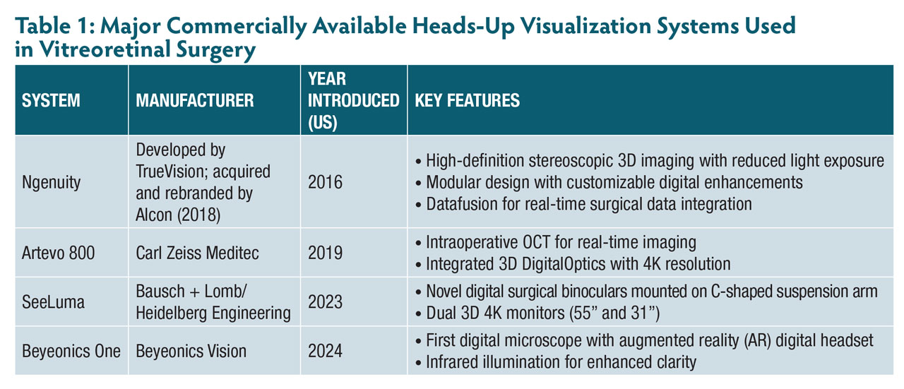
Over the past decade, heads-up surgical systems have rapidly evolved from a novel innovation to a tool used at academic institutions worldwide. Among currently available systems, Alcon’s Ngenuity platform, introduced in 2016, remains one of the most widely adopted. It offers high-definition 3D visualization via a 55-inch 4K monitor with customizable digital enhancements, such as color filters and tissue detail modes (Figure 2). Its modular design allows integration with existing analog microscope infrastructure, offering institutions flexibility in implementation. Since its release, systems like the Artevo 800 (Carl Zeiss Meditec) have further advanced the field. Introduced in 2019, the Artevo 800 was the first fully integrated digital microscope, combining hybrid optical-digital viewing with intraoperative OCT (iOCT) and image-guided overlays. Its ability to toggle between optical and digital views has made it especially valuable in educational settings.
Newer platforms continue to expand the capabilities of heads-up surgery (Table 1). The Beyeonics One system, introduced in 2024, offers a head-mounted display with voice control and immersive augmented reality (AR) features, reflecting a shift toward wearable and user-adaptable interfaces. Meanwhile, companies are exploring robotic integration and cloud-based data sharing. Looking ahead, advances may include AI-assisted intraoperative guidance, remote telesurgery, and virtual educational opportunities. As the field progresses, heads-up surgical systems may not only improve surgical precision and ergonomics but also fundamentally reshape the way vitreoretinal surgeons train, collaborate, and deliver care.
Challenges and Considerations
Although heads-up surgical systems offer numerous advantages, their integration into clinical practice is not without limitations. Institutions and surgeons must weigh financial, technical, and logistical considerations.
Fully integrated systems require substantial capital investment. Although modular platforms like Alcon’s Ngenuity offer flexibility for institutions with existing microscope infrastructure, there remain significant costs for installation, training, and periodic system upgrades. These financial barriers may be particularly restrictive for private practices and smaller surgical centers.
Furthermore, transitioning to heads-up visualization systems can disrupt established surgical workflows and often requires reconfiguring the operating room environment. Surgeons must adapt to changes in monitor positioning, lighting conditions, and ergonomics—for instance, positioning the screen over the patient’s legs or adjusting the iris aperture. Although many surgeons and trainees have praised heads-up systems for their educational value and enhanced visualization, others have reported reduced engagement during procedures, citing the bulky screen and the need for 3D glasses as drawbacks.
Intraoperatively, early users of heads-up systems have reported depth perception challenges, hand-eye coordination issues, and video latency. Peripheral visualization has also raised concern; however, related issues often stem from incorrect focus on the macula or excessive illumination, particularly in lightly pigmented fundi. Adjusting the light intensity and using color filters can enhance tissue detail and peripheral views.
Lastly, system compatibility and digital infrastructure are key considerations in the adoption of heads-up visualization systems. Some systems function best with specific microscopes, vitrectomy machines, or intraoperative OCT (iOCT) devices. Institutions must also ensure their infrastructure can support secure (HIPAA-compliant) data storage and remote consultation features. To implement these systems successfully, hospitals will need comprehensive training programs, robust institutional support, and careful workflow planning. Ultimately, the goal is to adopt technology that enhances surgical efficiency while safeguarding patient outcomes.
Conclusion
Heads-up surgical systems have introduced a new way of approaching visualization in the retinal operating room—one that prioritizes both patient outcomes and surgeon well-being. Over the past several years, platforms such as the Alcon Ngenuity and Zeiss Artevo 800 have demonstrated value in enhancing visualization, reducing endoillumination, and improving teaching environments. As these systems become more widely adopted, we may see that they offer more than just an ergonomic advantage—they may reshape how we interact with surgical anatomy, train the next generation of surgeons, and think about integrating digital tools into ophthalmic care.
Like most novel technologies, implementation comes with challenges that include cost, workflow integration, and learning curves. As this field continues to develop, heads-up surgical systems are well positioned to play a central role in the future of vitreoretinal surgery. RP


References
1. Baldwin G, Miller JB. Heads-up 3-dimensional visualization to enhance video endoscopy during vitreoretinal surgery. J Vitreoretin Dis. 2024;8(4):428-434. doi:10.1177/24741264241249527
2. Adam MK, Thornton S, Regillo CD, Park C, Ho AC, Hsu J. Minimal endoillumination levels and display luminous emittance during three-dimensional heads-up vitreoretinal surgery. Retina. 2017;37(9):1746-1749. doi:10.1097/IAE.0000000000001420
3. Baldwin G, Sokol JT, Ludwig CA, Miller JB. A comparative study of traditional scleral buckling to a new technique: guarded light pipe with heads-up three-dimensional visualization. Clin Ophthalmol. 2022; 16:3079-3088. doi:10.2147/OPTH.S378179
4. Moon JY, Seddon I, Sokol JT, et al. Comparison of ergonomics in vitreoretinal surgery with heads-up visualization versus the standard operating microscope as measured by a wearable device. Ophthalmic Surg Lasers Imaging Retina. 2024;55(11):638-645. doi:10.3928/23258160-20240508-02