



Proliferative vitreoretinopathy (PVR) remains the leading cause of failure after rhegmatogenous retinal detachment (RRD) repair, most commonly appearing 60 to 90 days after RRD. PVR occurs in approximately 5% to 10% of primary RRDs and represents an exaggerated wound-healing response following retinal break formation, retinal detachment, trauma, or retinal surgery. Major risk factors include large or multiple retinal breaks, giant retinal tears, chronic retinal detachment, extensive vitreous hemorrhage, choroidal detachment, ocular trauma, aphakia or pseudophakia, previous retinal surgery, uveitis, and preexisting PVR.1
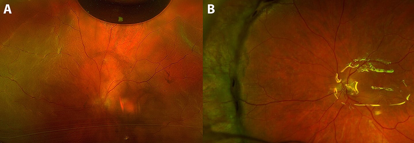
Figure 1. (A) Total retinal redetachment complicated by proliferative vitreoretinopathy, right eye, following pars plana vitrectomy (PPV) with laser and gas (bubble is present superior). (B) Retina reattachment following scleral buckle, PPV, laser, and silicone oil (note reflection of oil off macular region).
PVR Pathogenesis
PVR pathogenesis is a multistage process. The first phase involves blood-retinal barrier breakdown and cellular migration, which is initiated when a retinal tear occurs. This allows retinal pigment epithelium (RPE) cells, macrophages and other inflammatory cells, Müller cells, astrocytes, and fibroblasts to enter the vitreous cavity alongside serum proteins, altering the normal ocular environment and initiating an inflammatory cascade.
The resultant release of cytokines and growth factors (PDGF, TGF-β, VEGF, TNF-α, IL-1, IL-6, IL-8, and connective tissue growth factor) stimulates the next phase: cytokine signaling, phenotypic transformation, and further cellular proliferation, migration, and fibrosis. Under this signaling, RPE cells proliferate and undergo epithelial-mesenchymal transition (EMT) into contractile myofibroblast-like cells expressing α-smooth muscle actin (α-SMA).
Scaffold synthesis and retinal traction characterize the final phase. The transformed cells synthesize an abnormal extracellular matrix scaffold rich in collagen types I and II, fibronectin, and laminin, resulting in extracellular matrix contraction (EMC). Deposited above, below, and within the retina, this extracellular matrix causes retinal shortening and fixed folds (grade C PVR). This results in tangential and anteroposterior traction and subsequent recurrent retinal detachment.2
This pathogenesis can be addressed proactively through surgical and, potentially, pharmacologic PVR prophylaxis.
Surgical PVR Prophylaxis
Traditionally, surgical prophylaxis has been the primary defense, with techniques aimed at reducing liberated RPE cells and clearing inflammatory debris that drive PVR development. These include meticulous vitrectomy with removal of the posterior vitreous cortex, membrane peeling, relief of traction on retinal tears, and shaving of the vitreous base to minimize the physical scaffold required for cellular proliferation. Avoidance of cryotherapy, which can cause RPE dispersion and further blood-retinal barrier breakdown, may reduce stimuli for PVR development. The use of long-acting vitreous substitutes to maintain tear apposition and suppress membrane contraction, potential scleral buckling to support residual vitreous base traction on retinal tears, and, if necessary, relaxing retinotomy or circumferential (inferior) retinectomy to counteract retinal shortening are additional surgical maneuvers that may mitigate PVR.
Investigational Pharmacologic Adjuvants for PVR Prophylaxis
Investigational pharmacologic adjuvants for PVR prophylaxis target various phases of the PVR cascade, including inflammation, EMT, cell migration, and EMC. No agent has yet received regulatory approval for PVR prophylaxis or treatment. Studied agents include corticosteroids, which suppress inflammatory cytokines and cell migration; combined low-molecular-weight heparin and 5-fluorouracil, which inhibit cell proliferation and EMC; daunorubicin, which inhibits topoisomerase II and cell proliferation; infliximab, which inhibits TNF-α and EMT; dasatinib, a tyrosine kinase inhibitor that inhibits RPE and Müller cell growth, cell migration, EMT, and EMC; and Rho-associated protein kinase (ROCK) inhibitors, which inhibit cell migration, EMT, and EMC.3
Methotrexate (MTX) has been extensively studied for PVR prophylaxis. While high doses of MTX are used in oncology to disrupt DNA synthesis via folate antagonism, low doses act primarily as an anti-inflammatory agent. This low-dose pathway is mediated by increased extracellular adenosine (through inhibition of AICAR transformylase), which in turn suppresses inflammatory cytokines (TNF-α, IL-1, and IL-6), upregulates anti-inflammatory IL-10, induces immune cell apoptosis, and decreases neutrophil adhesion and aggregation.
Both intraoperative MTX infusions (40 mg of MTX dissolved in 500 mL of balanced salt solution) and intravitreal injections have been investigated. The standard intravitreal dose of MTX (400 µg) maintains therapeutic levels (0.1-2 µg/mL) for only 72 hours, necessitating frequent dosing (every 2 weeks), which can challenge patient adherence. To address this burden, sustained-release preparations are currently under development.
MTX has shown promise in the prophylaxis and treatment of PVR across recent clinical studies. A meta-analysis suggested that reoperation rates for RRD were significantly lower in eyes that received MTX at initial surgery compared with eyes that did not, although the analysis was limited by heterogeneity in treatment regimens across the 10 included studies.4 Furthermore, the Gain Understanding Against Retinal Detachment (GUARD) trial evaluated patients who received postoperative MTX injections following recurrent RRD due to PVR characterized by more than 3 clock hours of star folds or open-globe injury. This cohort experienced significantly fewer recurrent retinal detachments requiring reoperation within 6 months compared with a historical control group.5
Controversy
Continued refinements in vitreoretinal surgery, including ultrahigh-speed vitrectomy cutters, small-gauge instrumentation, and improved internal tamponades, have radically improved primary reattachment rates. Surgical maneuvers alone, however, cannot fully suppress an intrinsically cellular process. Surgical prophylaxis removes the structural scaffold but does not deactivate residual cellular activity or the inflammatory cytokines remaining within the ocular microenvironment. MTX may address this, but the logistical challenges of a multiple-injection protocol, potential MTX-related ocular surface toxicity, and the need for more definitive evidence of efficacy have limited its widespread use.
Given the available evidence and our individual experiences, should we rely on continued improvements in surgical PVR prophylaxis alone, or does the evidence supporting MTX warrant its use in most patients, or selected patients, undergoing RRD repair? Should adjunctive MTX become standard treatment in patients undergoing surgery for existing PVR or open-globe injuries? Are there other conditions, such as tractional retinal detachments that share downstream EMT mechanisms with PVR, that would warrant consideration of MTX?
We are fortunate to have commentaries addressing these questions from Vivienne S. Hau, MD, PhD, FASRS; John B. Miller, MD, with Ioanna Ploumi, MSc; and Christopher Riemann, MD, with Muhammad Jehanzeb Khan, MBBS, in this edition of Controversies in Care.
Beyond Surgical Success: Does Methotrexate Have a Definitive Role in PVR Prophylaxis?
Vivienne S. Hau, MD, PhD, FASRS
Every retina surgeon carries a few eyes they cannot forget. Mine belongs to a young man who came in with an open-globe injury. The repair went beautifully. He redetached anyway, star folds creeping across a retina I was sure I had saved. It is the case I picture when I read Dr. Colucciello's question: Does MTX have a definitive role in preventing PVR? My answer: It has earned a place in eyes like his, but I am not convinced it belongs in every detachment we repair.
The mechanics have improved, and sometimes that still isn't enough. Ultrahigh-speed cutters, small-gauge platforms, wide-angle viewing, and careful vitreous base dissection have all pushed my primary reattachment rates higher. But surgery works on the scaffold. It does not address the cellular and cytokine biology that continues for 6 to 8 weeks after we close.6 In an eye with grade C PVR, a giant retinal tear, a chronic macula-off detachment, or an open-globe injury, that proliferative drive is the very thing my instruments cannot reach. Those are the eyes in which a drug that quiets the biology makes sense to me.
What the strongest data say, and what they don’t. The signal for MTX is there, but it is not definitive, and I would rather say so plainly. The phase 3 GUARD trial showed a lower redetachment rate and encouraging secondary endpoints at 6 months after vitrectomy for PVR or open-globe injury, though the analysis relied partly on a historical control.6,7
A 2025 systematic review reported reattachment rates of 74% to 92% and fewer reoperations, though the numbers were small and the studies varied widely.4 The randomized data are what temper my enthusiasm. A multicenter randomized controlled trial (RCT) of intra-silicone oil MTX in grade C PVR did not reduce complete redetachment, although it did reduce membrane reproliferation and macular pucker.8 A 2025 meta-analysis of 3 RCTs involving 145 eyes found a 36% reduction in recurrence that did not reach statistical significance (relative risk, 0.64; 95% CI, 0.30-1.38).9 A 2026 network meta-analysis concluded that the evidence still does not support routine pharmacologic adjuvants, while identifying MTX as one of the more promising options.10 The direction is consistent, and the mechanism is attractive. What we lack is an adequately powered trial that demonstrates a statistically significant benefit for the outcome I care about: a retina that stays attached without a second trip to the operating room.
Should adjunctive MTX be standard for existing PVR or open-globe injuries? I think it is defensible. I would not yet call it mandatory. I offer serial MTX to eyes with recurrent PVR-associated detachment and to high-risk open-globe injuries, for which retrospective series report roughly 90% single-operation reattachment.11 I do not use it in routine primary RRD, where success rates are already high and the treatment burden is hard to justify. And it is a burden. The classic protocol can involve up to 13 injections, and corneal epitheliopathy occurs in approximately 15% to 33% of eyes. It is usually mild and reversible with longer dosing intervals and can be managed with folinic or folic acid.7,11,12 I walk patients through that trade-off the same way I would want it explained to me.
Looking beyond PVR. The epithelial-mesenchymal transition that MTX targets is not unique to PVR, so it is reasonable to wonder whether tractional retinal detachments and severe periretinal proliferation might benefit as well. mTOR inhibition is being explored on the same rationale.13 That is a hypothesis I want tested, not one I am ready to act on.
So this is where I have settled. I reach for MTX in the eyes most likely to fail me: recurrent PVR, established grade C disease, and high-risk open-globe injuries. I tell those patients that the definitive proof is not in yet. Sustained-release formulations that shorten the treatment schedule, together with the randomized primary RRD data that FIXER and similar trials will deliver, are what will move MTX from a reasonable option in selected eyes to standard of care. Until then, I try not to adopt it reflexively or dismiss it reflexively. And I keep thinking about that young man's eye.
The Role of MTX in PVR Management Remains Unclear
Ioanna Ploumi, MSc • John B. Miller, MD
The rationale for using MTX as an adjunct for PVR is compelling. Its antiproliferative and anti-inflammatory properties target important components of PVR pathogenesis. However, clinical studies evaluating its efficacy have yielded mixed results. The GUARD trial evaluated an intensive intravitreal MTX regimen, but its primary efficacy endpoint was compared with pooled historical controls.14 Concerns remain about the GUARD trial's historical control group because the numbers of eyes in the cited studies do not add up to the reported historical control totals. Furthermore, one of the included studies was published in 2007 and used vitreoretinal instrumentation that was less advanced than that used in the GUARD study nearly 2 decades later. The GUARD trial results have not been published in a peer-reviewed journal. This lack of a well-matched surgical control group prompted us to examine MTX use at our tertiary referral center, which has both high MTX utilization and extensive surgical experience with complex PVR detachments.
Our recent study at Massachusetts Eye and Ear evaluated 136 eyes undergoing repair of complex grade C PVR-related RDs, including 52 treated with adjunctive intravitreal MTX and 84 controls managed with advanced vitreoretinal surgical techniques without MTX. We found no significant difference in single-surgery retinal reattachment at 3 to 5 months, 1 year, or final follow-up. Increasing MTX exposure was also not associated with improved odds of attachment. Even among eyes receiving 13 or more injections, approximating the intensive regimen used in the GUARD trial, anatomic outcomes were not significantly different from those of controls.15
These findings should not be interpreted as evidence that MTX has no role in PVR. Rather, they raise what we believe is the more relevant question: Which patients, if any, derive additional benefit from MTX beyond optimized vitreoretinal surgery?
PVR is not a uniform disease. Eyes classified as grade C may differ considerably in the extent and location of PVR, inflammatory activity, chronicity, prior surgical history, and underlying etiology. It seems unlikely that every patient would derive the same benefit from adjunctive pharmacotherapy. Indeed, we agree with a central point raised in the subsequent discussion of our study: Not every patient with PVR necessarily needs MTX.16 At present, however, we lack validated clinical, imaging, or molecular biomarkers that can identify the subgroup most likely to benefit.
Beyond appropriate patient selection, we should also consider which surgeons may benefit most from MTX use. Advanced surgical techniques and the well-known “surgeon factor” in clinical studies can significantly influence the results of any recent or future MTX study. It would be possible to identify a surgeon or group of surgeons with poorer anatomic outcomes using advanced vitreoretinal techniques in the control group, making it more likely that the addition of MTX would demonstrate a statistically significant benefit. In their own practices, these surgeons might see greater benefit from MTX or another adjunctive therapy for complex PVR detachments. Alternatively, they could consider referring patients to surgeons with higher reattachment rates without MTX in complex PVR-related RD, when travel logistics permit. We observed similar effects of the surgeon factor in the PRO study results.
If MTX is chosen as adjunctive therapy, there is also considerable heterogeneity in how it is currently used. Published studies vary in dose, timing, frequency, and duration of treatment.17-20 Some use intraoperative MTX alone, whereas others use prolonged postoperative injection schedules.21,22 The optimal route of administration remains uncertain. This variability makes comparisons across studies difficult. The question, therefore, may not simply be whether MTX works, but in whom, when, and how it should be administered.
Recent evidence continues to reflect this uncertainty. Systematic reviews and meta-analyses have identified encouraging signals but have not demonstrated a consistent, statistically significant reduction in recurrent retinal detachment with MTX, and the overall certainty of evidence remains limited.9,10 Conversely, a recent small prospective randomized study of 50 eyes with grade C PVR reported improved single-surgery anatomic success with intraoperative MTX followed by 4 weekly injections.²³ This is encouraging, but the findings need confirmation in larger prospective trials.
It is also important to consider what MTX adds to contemporary PVR surgery. Advanced vitreoretinal techniques, including meticulous membrane peeling, appropriately extensive retinectomy or retinotomy, scleral buckling when indicated, and effective tamponade, can achieve favorable anatomic outcomes even in established grade C PVR.¹⁵ Favorable outcomes with an adjunctive therapy are most informative when compared with an appropriate control group, particularly because much of the published MTX literature consists of small or uncontrolled series.11,17,21
MTX also comes at a cost. Serial intravitreal injections increase treatment burden, cost, and postoperative visits following already complex retinal surgery. Corneal epitheliopathy is also a recognized adverse effect.24These considerations become particularly relevant when MTX is used broadly in patients who may already have favorable outcomes with surgery alone.
So, does MTX have a definitive role in PVR prophylaxis today? Probably not yet. This does not mean that MTX should be abandoned. There may well be selected eyes at particularly high risk for recurrent PVR in which adjunctive pharmacotherapy provides meaningful benefit. These include complex PVR-related RDs in pediatric patients and eyes with uveitic or traumatic etiologies. Current evidence does not support widespread routine use of MTX for every eye with established PVR or every RD considered at risk for PVR.
Rather than simply expanding its use, the next step should be to better define its role through prospective controlled studies with standardized surgical techniques and MTX protocols, careful PVR phenotyping, and, ultimately, biomarkers that may help identify treatment-responsive patients. For now, MTX remains a promising adjunct, but its optimal indications and definitive place in PVR management have yet to be established.
In Established PVR, the Signal Is Too Strong to Withhold
Christopher D. Riemann, MD • Muhammad Jehanzeb Khan, MBBS
Dr. Colucciello asks whether surgery alone is enough. In our experience, it is not. Vitrectomy removes the scaffold, but it cannot switch off the cells and cytokines left behind, and once PVR causes redetachment, only 40% to 80% of eyes recover ambulatory vision.2 Our quaternary care referral center has an extremely high and severe pathology burden and cares for profoundly unfortunate patients who have been referred in by other local retinal specialists or sometimes have been written off by other, far removed, centers of excellence. We have used MTX for PVR since our first patient in 2006, and our answer has 2 parts. In established PVR, the signal is now so strong that we cannot justify withholding MTX, even without level I evidence. In primary detachment, the question is still open, and we are answering it with a randomized trial.
We now treat every patient with established PVR. The basis is our 300-eye comparative cohort of retinal detachments with grade C PVR. Eyes treated with the Cincinnati MTX Protocol, a 40-mg/500-mL intraoperative infusion followed by 400-µg injections at postoperative weeks 1, 3, 6, and 10, redetached after a single surgery in 9.3% of cases, compared with 34.7% of untreated eyes (odds ratio for single-surgery success, 5.15; 95% CI, 2.77-10.15). Postoperative PVR decreased from 43% to 10%, and visual acuity was better at every time point through 12 months.25 The treated eyes had more severe disease, with more recurrent detachments (79% vs 55%), which biased the comparison against MTX. The association held in every sensitivity analysis, and an unmeasured confounder would need a risk ratio of nearly 4 to counter it. These are retrospective data from our own institution, but they agree with a separate 255-eye series in which postoperative MTX was associated with fewer than half as many reoperations as historical controls.26
The introduction also asks about tractional retinal detachment. Fibrovascular proliferation in diabetic retinopathy converges on the same fibrotic pathways, and in our cohort, none of the 15 treated eyes with proliferative diabetic retinopathy redetached, compared with 29% of untreated eyes.27 The 255-eye series included eyes with diabetic retinopathy and trauma, and open-globe injury was an entry criterion for GUARD.5,26 These eyes deserve the same consideration.
The 2 practical objections, injection burden and ocular surface toxicity, are real but manageable. As the introduction notes, a single injection maintains therapeutic levels for only about 72 hours, which is why the only regimen with phase 3 data requires 13 injections over 16 weeks.5 Our protocol front-loads the drug through the infusion, which adds no visit or needle, and schedules 4 injections during routine postoperative visits. In the grade C PVR cohort, 91% of eyes completed the series, and only 1 eye had injections withheld because of epitheliopathy, which resolved.25 MTX costs a few dollars per dose.27
Primary detachment is different. Most of these eyes will never develop PVR, so every patient treated carries the burden for a benefit only some will receive. That is the setting for FIXER (NCT06541574), which randomizes adults undergoing vitrectomy for primary RRD to MTX infusion, postoperative injections, both, or neither, with sham infusion and sham injections keeping patients and surgeons masked.28 At a prespecified interim analysis of the first 107 patients through week 10, no eye receiving both infusion and injections required reoperation, compared with approximately 1 in 4 controls. Each MTX arm outperformed sham, and there were no cases of endophthalmitis.27,29 That analysis was designed for safety, not efficacy, and we offer it as a signal rather than a definitive result.
The broader literature is less settled than our own experience. GUARD met its primary endpoint against historical controls, but in its smaller very underpowered concurrent comparison, reoperation for recurrent detachment occurred in 18.8% of treated eyes and 20.6% of controls.5,30 The meta-analysis cited in the introduction was favorable, but 2 meta-analyses published in 2026 found no statistically significant benefit albeit with significantly lower MTX dosing regimens.9,10 These studies mix populations, doses, and schedules, and none was powered for prevention in primary detachment. They show that the definitive trial has not been completed, and not that MTX fails.
Our answer, then, is 1) yes for established PVR, 2) yes to PDR, 3) yes in eyes with primary RD and elevated risk for PVR such as posterior uveitis, trauma and a history of contralateral PVR for which we believe MTX should be standard, and 4) not yet for uncomplicated primary detachment, for which we randomize. We have expanded FIXER beyond our institution, with masked adjudication and unplanned reoperation as the primary outcome. Please consider joining this important labour of love by becoming a study site for FIXER. MTX is not yet proven for every detachment, but it is closer than any agent before it, and the trial that can settle the question is underway. RP





REFERENCES
1. Lin JB, Wu F, Kim LA. Proliferative vitreoretinopathy: pathophysiology and therapeutic approaches. Int Ophthalmol Clin. 2024;64(2):125-135. doi:10.1097/IIO.0000000000000495
2. Idrees S, Sridhar J, Kuriyan AE. Proliferative vitreoretinopathy: a review. Int Ophthalmol Clin. 2019;59(1):221-240. doi:10.1097/IIO.0000000000000258
3. Khosravi Mirzaei S, Hatami F, Safi S, et al. Pharmacological agents for treatment of proliferative vitreoretinopathy: a systematic review and network meta-analysis. Surv Ophthalmol. 2025;70(4):633-644. doi:10.1016/j.survophthal.2025.02.005
4. Toh VTR, Gerard G, Tay ZQ, Chen J, Chew GWM, Teoh CS. Efficacy and safety of methotrexate in the treatment of proliferative vitreoretinopathy: a systematic review. Eye (Lond). 2025;39(3):460-467. doi:10.1038/s41433-024-03503-55
5. The GUARD trial, part 1: a phase 3 clinical trial for prevention of proliferative vitreoretinopathy. Clinicaltrials.gov identifier: NCT04136366. Updated July 9, 2025. Accessed October 16, 2026. https://clinicaltrials.gov/study/NCT04136366
6. Ferro Desideri L, Artemiev D, Zandi S, Zinkernagel MS, Anguita R. Proliferative vitreoretinopathy: an update on the current and emerging treatment options. Graefes Arch Clin Exp Ophthalmol. 2024;262(3):679-687. doi:10.1007/s00417-023-06264-1
7. Wang WX, Xing M, Apte RS. Interventions for proliferative vitreoretinopathy. JAMA Ophthalmol. 2024;142(7):669-670. doi:10.1001/jamaophthalmol.2024.1093
8. Nourinia R, Safi S, Mohammadpour M, et al. Effects of repeated intra-silicone oil injections of methotrexate on proliferative vitreoretinopathy grade C: a multicenter randomized controlled trial. Sci Rep. 2024;14(1):28842. doi:10.1038/s41598-024-79708-1
9. Kocak I, Sayin N, Bayramoglu SE, Thalib L. Effect of methotrexate in preventing retinal detachment after proliferative vitreoretinopathy surgery: a systematic review and meta-analysis. Surv Ophthalmol. 2026;71(2):572-581. doi:10.1016/j.survophthal.2025.09.015
10. Al-Burak SA, Alizada A, Sadek K, Aleshawi A, Al-Dwairi R, Abu Serhan H. Pharmacologic interventions for the prevention and treatment of proliferative vitreoretinopathy: a systematic review and meta-analysis. Ophthalmol Retina. Published online August 7, 2026:S2468-6530(26)00391-X. doi:10.1016/j.oret.2026.08.002
11. Hughes PJ, Bhagat N, Gonzalez-Martinez OG, Zarbin MA. Intravitreal methotrexate injection for the treatment and prevention of proliferative vitreoretinopathy. Retina 2024;44(10):1748-1757. doi:10.1097/IAE.0000000000004181
12. Gorovoy I, Prechanond T, Abia M, Afshar AR, Stewart JM. Toxic corneal epitheliopathy after intravitreal methotrexate and its treatment with oral folic acid. Cornea. 2013;32(8):1171-1173. doi:10.1097/ICO.0b013e3182910106
13. Alsoudi AF, Loya A, Wai K, et al. Mammalian target rapamycin inhibition as a therapeutic target for prevention of proliferative vitreoretinopathy. JAMA Ophthalmol. 2025;143(9):777-784. doi:10.1001/jamaophthalmol.2025.2497
14. Aldeyra Therapeutics completes enrollment in part 1 of the phase 3 GUARD trial of ADX2191 in proliferative vitreoretinopathy. News release. January 4, 2022. Accessed October 16, 2026. https://ir.aldeyra.com/node/11231/pdf
15. Ploumi I, Garcia MD, Zhu Y, et al. Outcomes of complex PVR retinal detachment repair with and without intravitreal methotrexate at a single tertiary care center. Ophthalmic Surg Lasers Imaging Retina. 2026;57(2):83-92. doi:10.3928/23258160-20250826-01
16. Ploumi I, Miller JB. Reply to letter to the editor: intravitreal methotrexate for complex retinal detachment. Ophthalmic Surg Lasers Imaging Retina. 2026;57(4):269-271. doi:10.3928/23258160-20260309-01
17. Benner JD, Dao D, Butler JW, Hamill KI. Intravitreal methotrexate for the treatment of proliferative vitreoretinopathy. BMJ Open Ophthalmol. 2019;4(1):e000293. doi:10.1136/bmjophth-2019-000293
18. Nourinia R, Borna F, Rahimi A, et al. Repeated injection of methotrexate into silicone oil–filled eyes for grade C proliferative vitreoretinopathy: a pilot study. Ophthalmologica. 2019;242(2):113-117. doi:10.1159/000500271
19. Liu T, Runner M, Mahmoud TH, et al. A Modified Intravitreal Methotrexate Protocol for the Prevention of Proliferative Vitreoretinopathy after Silicone Oil Removal. Ophthalmol Retina. 2025;9(4):314-321. doi:10.1016/j.oret.2024.10.006
20. Ullah A, Toth CA, Burnett HW, Butler JW, Levy JH, Benner JD. Low-dose intravitreal methotrexate for proliferative vitreoretinopathy. Ophthalmic Surg Lasers Imaging Retina. 2023;54(3):139-146. doi:10.3928/23258160-20230220-01
21. Sadaka A, Sisk RA, Osher JM, Toygar O, Duncan MK, Riemann CD. Intravitreal methotrexate infusion for proliferative vitreoretinopathy. Clin Ophthalmol. 2016;10:18111817. doi:10.2147/OPTH.S111893
22. Jahangir S, Jahangir T, Ali MH, Lateef Q, Hamza U, Tayyab H. Use of intravitreal methotrexate infusion in complicated retinal detachment for prevention of proliferative vitreoretinopathy in a pilot study. Cureus. 2021;13(8):e17439. doi:10.7759/cureus.17439
23. Mohamed Elsayed Saad E, Elbadry Mohammed Mohammed H, Dabour SA, Alyan A. Methotrexate as an adjunct to pars plana vitrectomy for the prevention and treatment of proliferative vitreoretinopathy in rhegmatogenous retinal detachment: a clinical study. Graefes Arch Clin Exp Ophthalmol. Published online August 26, 2026. doi:10.1007/s00417-026-07467-y
24. Shen-Sampas JH, Ahmad TR, Stewart JM. Corneal toxicity of intravitreal methotrexate used for the treatment of proliferative vitreoretinopathy in silicone oil–filled eyes: a case series. Cornea. 2022;41(4):499-501. doi:10.1097/ICO.0000000000002774
25. Huang D, Feist JE, McTavish SA, et al. Outcomes of Complex Retinal Detachment Repair with Proliferative Vitreoretinopathy Grade C using the Cincinnati Methotrexate Protocol Ophthalmol Retina. Published online October 14, 2026. doi:10.1016/j.oret.2026.09.030
26. Gibson L, Nguyen A, Ridgeway J, et al. Postoperative Methotrexate to Reduce Reoperation Rate and Improve Vision in Patients With Complex Retinal Detachments, Advanced Diabetic Retinopathy, and Trauma. J Vitreoretin Dis. 2025;9(3):336-342. doi:10.1177/24741264251323305
27. Franklin AJ, Gibson Snellings L, Nguyen A, Riemann CD. Methotrexate: an emerging surgical adjuvant. Retina Today. January/February 2026. Accessed October 16, 2026. https://retinatoday.com/articles/2026-jan-feb/methotrexate-an-emerging-surgical-adjuvant
28. Prevention of ProliFerative Vitreoretinopathy with Intravitreal MethotreXate in Primary Retinal DEtachment Repair (FIXER) trial. Clinicaltrials.gov identifier: NCT06541574. Updated September 19, 2024. Accessed October 16, 2026. https://clinicaltrials.gov/study/NCT06541574
29. Riemann CD. Interim report of the FIXER trial. Presented at: ASRS Annual Meeting; August 2, 2025; Long Beach, CA.
30. Aldeyra Therapeutics achieves primary endpoint in Part 1 of Phase 3 GUARD trial of ADX-2191 in proliferative vitreoretinopathy. Press release. October 6, 2022. Accessed October 16, 2026. https://ir.aldeyra.com/news-releases/news-release-details/aldeyra-therapeutics-achieves-primary-endpoint-part-1-phase-3