



For decades, retinal imaging has steadily improved physicians' ability to visualize disease. Yet even the highest-resolution structural images cannot answer a fundamental question: Are the photoreceptors still functioning normally? That gap, argued Kareem Moussa, MD, is where optoretinography (ORG) may find its role.
Presenting early clinical experience with the technology at the American Society of Retina Specialists (ASRS) annual meeting in Montreal, Dr. Moussa, a retina specialist at the University of California, Davis, working in collaboration with vision scientists Ravi Jonnal, PhD, and Robert Zawadzki, PhD, described ORG as a functional imaging technique that measures photoreceptor responses to light using a standard OCT light source.
“ORG is an emerging technology that I think is going to change your life,” he told attendees Thursday.
Current clinical decision-making relies primarily on structural findings, Dr. Moussa said. Retina specialists identify edema, leakage, or other anatomic abnormalities and determine treatment accordingly. Functional testing has remained on the periphery because available tools have important limitations.
“We don't really consider function,” he said. “It’s not because we don’t want to. It’s because the tests that we currently have to assess function are not very good."
ORG seeks to address that limitation by capturing subtle movements of photoreceptor outer segments after light stimulation. Following exposure to the stimulus, photoreceptors undergo an initial contraction followed by elongation. ORG quantifies the photoreceptor response using 2 velocity-based metrics: Vmin, which measures the initial contraction immediately after light stimulation, and V20-40, which measures the average rate of photoreceptor elongation 20 to 40 milliseconds later.
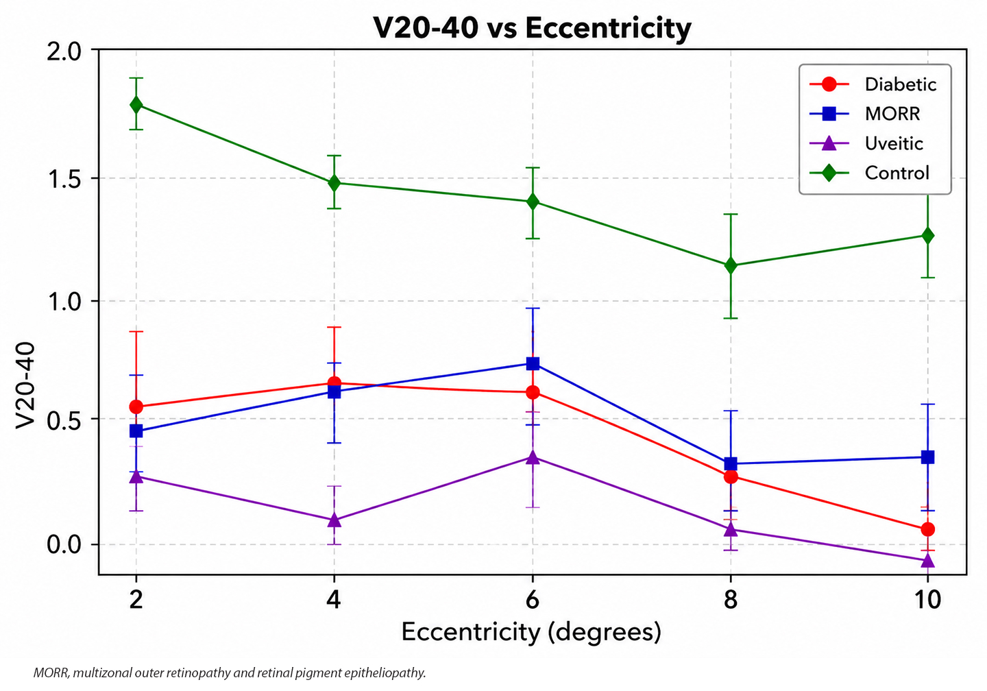
Dr. Moussa presented findings from a prospective UC Davis study that included healthy eyes and eyes with retinal disease, primarily diabetic retinopathy and uveitis. Analysis of the V20-40 metric showed a consistent reduction in photoreceptor elongation among diseased eyes compared with healthy controls. In normal eyes, elongation velocity slowed with increasing distance from the fovea, whereas eyes with retinal disease demonstrated attenuation across the measured retinal locations (Figure 1).
The investigators then examined a more clinically challenging population: patients with diabetes who had no evidence of diabetic retinopathy on structural examination. “These are patients who you'd see in the clinic and say, ‘Your eyes are fine. Come back in a year,’” noted Dr. Moussa. However, even in those structurally normal eyes, ORG detected evidence of altered photoreceptor function compared with healthy controls, suggesting that functional abnormalities may precede visible structural changes.
“In summary, with ORG we’re able to measure photoreceptor contraction and elongation, and we’re able to detect differences between eyes that have disease and eyes that are normal,” Dr. Moussa said. He cautioned that the technology remains in an early stage of development: “We still have quite a bit more work to do to validate this further.” RP