


For retina specialists, understanding neuro-ophthalmology is essential because the eye and brain are closely connected, and some patients present with symptoms that could stem from either a retinal disease or a neurologic disorder. Andrew G. Lee, MD, urged retinal physicians to sharpen their diagnostic skills at this intersection of the eye and the brain during a presentation Saturday at the 2025 American Academy of Ophthalmology (AAO) meeting in Orlando, Florida.
“Neuro-ophthalmology and retina patients overlap, and we meet at the optic nerve,” said Dr. Lee, chair of the Blanton Eye Institute at Houston Methodist Hospital in Houston, Texas. “Sometimes it’s not clear to retina doctors when it’s retina and when it’s neuro-ophthalmology.”
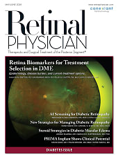
Neuro-Op Pearls for Retina
- A stroke of the eye (CRAO) is a stroke
- Optic atrophy does not necessarily mean optic nerve pathology (BRAO, CRAO)
- Optic atrophy does not occur in CME, AMD, macular hole, ERM, PVD, CRNVM
- Test visual field in all unexplained visual loss
- Test peripheral field in every patient (confrontation minimum)
- Big blind spot is only a neuro-op VF defect if papilledema
- Vertical step in visual field is neuro-op
- MRI head/orbit, gadolinium, fat sat for any optic neuropathy
In his retina subspecialty day talk, Dr. Lee noted that retina specialists must identify when an apparent retinal disorder may actually originate from the optic nerve or elsewhere in the nervous system. In such cases, additional testing, including electrophysiology, may be required to confirm a retinal cause. Conversely, when the retina appears abnormal but the underlying pathology is neuro-ophthalmic—such as inflammation or tumor involvement—misdiagnosis can delay appropriate care.
“The short answer is, you should image a retina case when it’s a special kind of retina, called ‘not the retina,’” Dr. Lee explained. “The trick is determining when it is ‘not the retina,’ even though it looks like the retina initially may be the problem.” He stressed that careful attention to clinical clues can guide decision-making. For example, a central scotoma often indicates optic neuropathy, while a ring scotoma or big blind spot (without papilledema) with photopsias suggest retinal disease.
Other red flags include light-dependent visual disturbances. “The photoreceptors care about light and dark, but the optic nerve really doesn’t care,” Dr. Lee said. “So, things like night blindness (nyctanopia) and day blindness (hemeranopia)—these are retina problems, but the retina in these retinopathy cases sometimes looks normal or near normal initially.” In some patients, these presentations may reflect cancer-associated retinopathies or other paraneoplastic syndromes, highlighting the need for systemic evaluation.
Dr. Lee advised retina specialists to adopt several neuro-ophthalmologic assessments as routine to help differentiate the conditions. “You should be doing some of the things that we in neuro-op always do,” he said. “Swing the flashlight for a relative afferent pupillary defect (RAPD), check the color vision, do a formal visual field (looking for a vertical step), and then perform the OCT of both the optic nerve and the macula.” These tools, he noted, can help distinguish retinal from optic nerve pathology (eg, optic atrophy) without adding significant workflow burden.
Imaging, when indicated, requires precision in ordering. “If you’re going to the trouble of ordering an MRI for an optic neuropathy then it has to be ‘MR head, orbit, gadolinium, fat sat,’” Dr. Lee said. Inclusion of both head and orbit ensures visualization of intracranial and intraorbital portions of the optic nerve; gadolinium highlights inflammatory or neoplastic lesions; and fat suppression prevents orbital fat from obscuring pathology.
The diagnostic overlap also extends to vascular disease, because the paradigm for ocular strokes has shifted, he observed. Branch or central retinal artery occlusions, once managed as isolated ophthalmic events, are now recognized as strokes requiring emergency evaluation. Patients with acute retinal arterial occlusions must be referred directly to stroke centers within the 4.5-hour to 6-hour treatment window. “The thing that’s changed for retina doctors is recognizing that a stroke in the eye is a stroke,” Dr. Lee said. “Time is brain and time is retina, and they need to go to the stroke center.”
The bottom line, said Lee, is that retina doctors need to know when the visual loss is the retina but also when it is “not the retina.” RP